
Giada Cucciolini12, Diego Costanzo1, Antonella Ghetta1, Francesco De Masi1
 ORCID
ORCID
Recibido: 2026-02-05
Aceptado: 2026-06-12
©2026 El(los) Autor(es) – Esta publicación es Órgano oficial de la Sociedad de Anestesiología de Chile
Revista Chilena de Anestesia Vol. 55 Núm. 5 | https://doi.org/10.25237/revchilanestv55n5-24
PDF
Utilidad de la ecografía a pie de cama en el manejo de la malposición de un catéter venoso central en cuidados neurocríticos: Reporte de un caso
Abstract
We report a case of a 36-year-old male admitted to the ICU for subarachnoid hemorrhage. Following external ventricular drain insertion and aneurysm embolization, a 4-lumen CVC was placed via the right subclavian vein under ultrasound (US) guidance. Post-procedural chest X-ray suggested malposition in the brachiocephalic vein. However, subsequent US re-evaluation in the supraclavicular window, combined with the insertion of a guidewire visualized in the right atrium, confirmed correct positioning at the cavoatrial junction. A follow-up X-ray confirmed the spontaneous repositioning. This case highlights the critical role of bedside ultrasound in verifying CVC tip location, preventing unnecessary and risky re-interventions and allows for thoughts about the rigidity of searching for the cavoatrial junction exact positioning. Ultrasound provides a dynamic assessment that can resolve discrepancies between static radiographic imaging and actual intravascular position, enhancing patient safety.
Resumen
We report a case of a 36-year-old male admitted to the ICU for subarachnoid hemorrhage. Following external ventricular drain insertion and aneurysm embolization, a 4-lumen CVC was placed via the right subclavian vein under ultrasound (US) guidance. Post-procedural chest X-ray suggested malposition in the brachiocephalic vein. However, subsequent US re-evaluation in the supraclavicular window, combined with the insertion of a guidewire visualized in the right atrium, confirmed correct positioning at the cavoatrial junction. A follow-up X-ray confirmed the spontaneous repositioning. This case highlights the critical role of bedside ultrasound in verifying CVC tip location, preventing unnecessary and risky re-interventions and allows for thoughts about the rigidity of searching for the cavoatrial junction exact positioning. Ultrasound provides a dynamic assessment that can resolve discrepancies between static radiographic imaging and actual intravascular position, enhancing patient safety.
Introduction
Central venous catheterization (CVC) is a fundamental procedure in the management of neurocritical care patients, providing essential access for hemodynamic monitoring, administration of hyperosmolar therapies, and vasoactive drugs. In the setting of subarachnoid hemorrhage (SAH), the choice of the insertion site is critical. The right subclavian vein is often preferred over the internal jugular vein to avoid potential interference with cerebral venous drainage-which could exacerbate intracranial hypertension-and to maintain a safe distance from eventual tracheostomy sites, thereby reducing the risk of catheter-related infections. Despite the widespread adoption of real-time ultrasound (US) guidance, which has significantly increased success rates and reduced immediate mechanical complications[1]-[3], catheter tip malposition remains a documented challenge. While chest X-ray (CXR) is traditionally considered the gold standard for confirming the final position of the tip at the cavoatrial junction, it provides only a static, two-dimensional projection that may occasionally lead to misinterpretation due to patient positioning or anatomical variations[4]. Recent literature emphasizes the role of Point-of-Care Ultrasound (POCUS) as a dynamic tool for tip navigation and location. However, discrepancies between initial radiographic findings and subsequent clinical assessments can occur, especially following patient mobilization or routine nursing care. This case report describes a 36-year-old patient with SAH where an apparent malposition on CXR was resolved spontaneously through a systematic ultrasound re-evaluation using the “wire-target” technique. This approach prevented an unnecessary and potentially risky re-intervention, highlighting the importance of multi-modal verification in the Intensive Care Unit (ICU).
Case description
A 36-year-old male was admitted to the Intensive Care Unit (ICU) following a subarachnoid hemorrhage (SAH) caused by a ruptured anterior communicating artery (ACoA) aneurysm. Upon arrival, the patient presented with a Glasgow Coma Scale (GCS) score of 14. Due to acute hydrocephalus, an external ventricular drain (EVD) was placed, followed by successful endovascular embolization of the aneurysm using coils. The patient was initially managed with mechanical ventilation under sedation but was subsequently extubated, maintaining a stable neurological status with a GCS of 14.
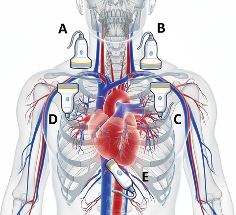
Figure 1. Probe positioning and US windows during positioning verification of the CVC. A: supraclavicular window, right side; B: supraclavicular window, side left; C: infraclavicular window, left side; D: infraclavicular window, right side, E: subcostal bicaval window.
On the third day of hospitalization, a decision was made to insert a 4-lumen central venous catheter (CVC). The right subclavian vein was selected as the insertion site, following neuro-ICU protocols to minimize interference with cerebral venous return and reduce the risk of infection compared to internal jugular or femoral access. The procedure was performed under real-time ultrasound guidance with an apparently uncomplicated insertion. The homolateral jugular vein was checked intraprocedurally and resulted empty.
After CVC positioning the patient underwent routine hygiene and mobilization. However, the post-procedural chest X-ray (CXR) revealed a malposition, with the catheter tip appearing to be lodged within the left brachiocephalic vein (Figure 1). Before proceeding with a potentially unnecessary catheter replacement, a comprehensive Point-of-Care Ultrasound (POCUS) assessment was performed to re-evaluate the catheter’s trajectory (Figure 1). The ultrasound examination yielded the following findings[1]: the check of the left brachiocephalic vein and subclavian vein evidenced the absence of any catheter (Figure 1, Position B). The left jugular vein was empty as well[2]. While observing the left brachiocephalic trunk, a flush test was performed: a distal port saline flush was not visible, suggesting that malpositioning in the left brachiocephalic trunk was improbable[3]. Visualization of the right supraclavicular window (Figure 1, Position A) allowed visualization of the right innominate vein and the confluence of the right internal jugular and subclavian veins. The junction of the two brachiocephalic trunk was visible as well and, in this view, the catheter was seen directed inferiorly toward the superior vena cava, suggesting a correct anatomical course despite the initial radiographic interpretation (Figure 2)[4].
To definitively confirm the tip location, a guidewire was advanced through the distal port of the CVC under continuous ultrasound monitoring. The wire was clearly visualized entering the right atrium (Figure 3). In addition, injection of contrast (blood + air + saline) was clearly visible coming out from the distal port next to the atrium (Figure 1, Position E). These maneuvers confirmed that the catheter tip had spontaneously repositioned in a correct, functional position at the cavoatrial junction. A follow-up CXR was then performed, which confirmed the definitive and correct positioning of the tip (Figure 1). The use of POCUS and the “wire-target” technique successfully prevented an invasive re-intervention and ensured patient comfort.
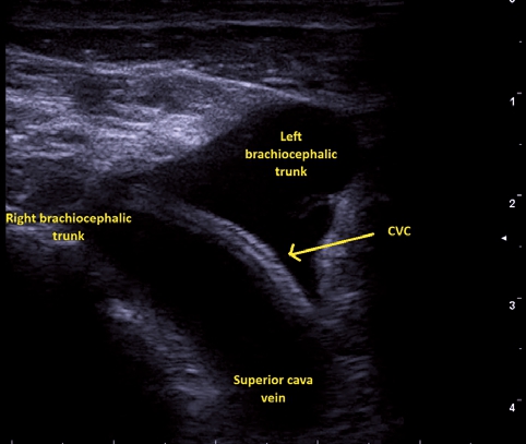
Figure 2. US image from the right upper supraclavicolar US window. CVC direction is clearly visible and directed towards the superior vena cava.
Discussion
The discrepancy between initial radiographic findings and the actual intravascular position of a central venous catheter (CVC) is a well-documented phenomenon that poses a clinical dilemma in the ICU[5]. While post-procedural chest X-ray (CXR) remains a traditional standard for tip verification, its reliability is limited by its two-dimensional projection, which can lead to “apparent” malpositions due to anatomical overlaps or patient rotation.
In our case, the initial CXR suggested a malposition in the left brachiocephalic vein. However, subsequent clinical management-specifically routine patient hygiene and mobilization-likely facilitated a spontaneous redirection of the catheter tip towards the cavoatrial junction. Literature suggests that movements of the neck and upper limbs, especially abduction and rotation, can induce significant tip migration (up to 2-3 cm) in catheters inserted via the subclavian route[6],[7]. This highlights that a “static” malposition on an early X-ray may resolve spontaneously with patient positioning or simple manoeuvres.
The use of Point-of-Care Ultrasound (POCUS) proved decisive in this scenario. While the standard infraclavicular view confirms the entry into the vein, the supraclavicular window allows for direct visualization of the confluence of the internal jugular and subclavian veins and the course of the brachiocephalic vein towards the superior vena cava. The “wire-target” technique we employed-advancing a guidewire under real-time ultrasound-provided definitive confirmation of the tip’s location within the right atrium.
Recent studies have shown that POCUS has a high sensitivity (up to 96%) and specificity for detecting CVC malpositions, often identifying correct placement where CXR might be ambiguous[5],[8]. Furthermore, ultrasound verification is significantly faster than waiting for radiographic confirmation, reducing the time to utilize the catheter for critical therapies[9].
The spontaneous repositioning of the CVC observed in our case necessarily implies that the catheter tip underwent a clinically significant displacement within the central venous vascular tree, likely driven by changes in venous flow, intrathoracic pressure, and vessel geometry related to respiration and patient positioning.
This dynamic behaviour challenges, at least from a practical standpoint, the traditional dogma that defines the cavo-atrial junction as a strictly fixed and definitive target for optimal tip placement[10]. Our local observation suggests that catheter tip location should not always be considered a static parameter, but rather a potentially variable one even in the absence of external manipulation.
This supports the concept that central venous anatomy and catheter position are dynamic, and it reinforces the value of repeatable bedside imaging methods, such as point-of-care ultrasound, for ongoing verification of tip location[4],[9],[10].
By integrating clinical re-evaluation with advanced POCUS techniques, we avoided an unnecessary second invasive procedure. This not only eliminated the inherent risks of a new venipuncture-such as pneumothorax or arterial injury-but also preserved patient comfort and reduced ICU costs. This case underscores that a multidisciplinary approach, combining radiology with dynamic ultrasound verification, should be the standard of care when facing suspected CVC malpositions.
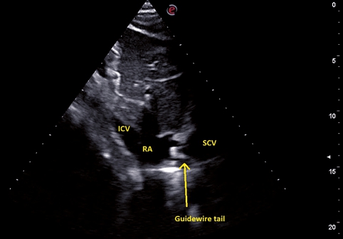
Figure 3. Bicaval subcostal window showing the presence of the guidewire tail at the cavoatrial junction.
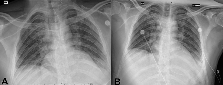
Figure 4. Chest X ray of the patient before (left side, panel A) and after (right side, panel B) CVC spontaneous repositioning.
Referencias
1. Brass P, Hellmich M, Kolodziej L, Schick G, Smith AF. Ultrasound guidance versus anatomical landmarks for internal jugular vein catheterization. Cochrane Database Syst Rev. 2015 Jan 9;2015(1):CD006962.
2. Vezzani A, Manca T, Brusasco C, Santori G, Cantadori L, Ramelli A, et al. A randomized clinical trial of ultrasound-guided infra-clavicular cannulation of the subclavian vein in cardiac surgical patients: short-axis versus long-axis approach. Intensive Care Med. 2017 Nov;43(11):1594-601. https://doi.org/10.1007/s00134-017-4756-6 PMID: https://pubmed.ncbi.nlm.nih.gov/28289815.
3. Sidoti A, Brogi E, Biancofiore G, Casagli S, Guarracino F, Malacarne P, et al. Ultrasoundversus landmark-guided subclavian vein catheterization: a prospective observational study from a tertiary referral hospital. Sci Rep. 2019 Aug 22;9(1):12248. https://doi.org/10.1038/s41598-019-48766-1 PMID: https://pubmed.ncbi.nlm.nih.gov/31439913.
4. Corradi F, Cucciolini G, Tavazzi G, Wong A, Balan C, Melniker LA, et al. WINFOCUS worldwide survey on central venous catheter insertion and position confirmation practices (CVC-ICON study). Ultrasound J. 2025 Aug 14;17(1): 41. https://doi.org/10.1186/s13089-025-00429-1 PMID: https://pubmed.ncbi.nlm.nih.gov/40810971.
5. Venugopal AN, Koshy RC, Koshy SM. Role of chest X-ray in citing central venous catheter tip: A few case reports with a brief review of the literature. J Anaesthesiol Clin Pharmacol. 2013 Jul;29(3):397-400.
6. Jahagirdar SM, Laxmimani, Athiraman U, Ravishankar M. Migration of subclavian venous catheter tip: Patient positioning in ICU makes a difference. Indian J Crit Care Med Peer-Rev Off Publ Indian Soc Crit Care Med. 2013;17(3):193-4. https://doi.org/10.4103/09725229.117085 PMID: https://pubmed.ncbi.nlm.nih.gov/24082622.
7. Rajan S, Paul J, Kumar L. Spontaneous repositioning of a malpositioned peripherally inserted central catheter. Indian J Anaesth. 2016 Feb;60(2):148-9. https://doi.org/10.4103/0019-5049.176283 PMID: https://pubmed.ncbi.nlm.nih.gov/27013762.
8. Vezzani A, Brusasco C, Palermo S, Launo C, Mergoni M, Corradi F. Ultrasound localization of central vein catheter and detection of postprocedural pneumothorax: an alternative to chest radiography. Crit Care Med. 2010 Feb;38(2):533-8. https://doi.org/10.1097/ccm.0b013e3181c0328f PMID: https://pubmed.ncbi.nlm.nih.gov/19829102.
9. de Man L, Wentzel M, van Rooyen C, Turton E. Comparison between ultrasound and chest X-ray to confirm central venous catheter tip position. SA J Radiol. 2023;27(1):2587. https://doi.org/10.4102/sajr.v27i1.2587 PMID: https://pubmed.ncbi.nlm.nih.gov/37416693.
10. Hill S, Moureau NL. Tip Position. In: Moureau NL, editor. Vessel Health and Preservation: The Right Approach for Vascular Access [Internet]. Cham: Springer International Publishing; 2019 [cited 2026 Feb 6]. p. 81-105. Available from: https://doi.org/10.1007/978-3-030-03149-7_7