
Saeid Elsawy1, Mostafa Abulfotouh1, Obay Elhasan Shaker1, Eman M. Khedr1, Rasha Hamed1, Yara Hamdy Abbas1
 ORCID
ORCID
Recibido: 2026-03-09
Aceptado: 2026-06-21
©2026 El(los) Autor(es) – Esta publicación es Órgano oficial de la Sociedad de Anestesiología de Chile
Revista Chilena de Anestesia Vol. 55 Núm. 5 | https://doi.org/10.25237/revchilanestv55n5-17
PDF
Eficacia de Navi-Cath versus catéter RACZ en el manejo del síndrome post-laminectomía: Un ensayo clínico prospectivo aleatorizado
Abstract
Background: Failed back surgery syndrome (FBSS) is a common cause of persistent lumbar and/or radicular pain following spinal surgery, often driven by epidural fibrosis. Percutaneous adhesiolysis (PA) has emerged as an effective treatment, yet the optimal catheter design remains unclear. This study compared the efficacy of the Racz catheter (non-steerable, smaller caliber) and the Navi-Cath catheter (steerable, larger caliber) in FBSS patients. Methods: Sixty patients with FBSS were randomized to receive adhesiolysis using either a Racz (n = 30) or Navi-Cath (n = 30) catheter. Pain intensity (Numeric Rating Scale, NRS), functional disability (Oswestry Disability Index, ODI), pregabalin use, and complications were assessed at baseline, 1, 3, 6, and 12 months. Results: Baseline characteristics were similar between groups. No significant differences were noted in NRS at 1 and 3 months. At 6 and 12 months, the Navi-Cath group reported significantly greater pain reduction (p = 0.01, p=0.002). ODI scores improved significantly in both groups, with consistently superior functional recovery in the Navi-Cath group (p < 0.001). Pregabalin use decreased in both groups, with lower but non-significant reductions in the Navi-Cath group. Complication rates were comparable. Conclusion: Navi-Cath adhesiolysis provided superior long-term pain relief and functional recovery compared with the Racz catheter, without increased complications. These findings suggest that steerable catheters may offer clinical advantages in FBSS management.
Resumen
Background: Failed back surgery syndrome (FBSS) is a common cause of persistent lumbar and/or radicular pain following spinal surgery, often driven by epidural fibrosis. Percutaneous adhesiolysis (PA) has emerged as an effective treatment, yet the optimal catheter design remains unclear. This study compared the efficacy of the Racz catheter (non-steerable, smaller caliber) and the Navi-Cath catheter (steerable, larger caliber) in FBSS patients. Methods: Sixty patients with FBSS were randomized to receive adhesiolysis using either a Racz (n = 30) or Navi-Cath (n = 30) catheter. Pain intensity (Numeric Rating Scale, NRS), functional disability (Oswestry Disability Index, ODI), pregabalin use, and complications were assessed at baseline, 1, 3, 6, and 12 months. Results: Baseline characteristics were similar between groups. No significant differences were noted in NRS at 1 and 3 months. At 6 and 12 months, the Navi-Cath group reported significantly greater pain reduction (p = 0.01, p=0.002). ODI scores improved significantly in both groups, with consistently superior functional recovery in the Navi-Cath group (p < 0.001). Pregabalin use decreased in both groups, with lower but non-significant reductions in the Navi-Cath group. Complication rates were comparable. Conclusion: Navi-Cath adhesiolysis provided superior long-term pain relief and functional recovery compared with the Racz catheter, without increased complications. These findings suggest that steerable catheters may offer clinical advantages in FBSS management.
Introduction
Failed back surgery syndrome (FBSS) is defined as persistent radicular and/or lumbar pain lasting beyond three months following spinal surgery (Barito et al., 2019; Lewik et al., 2023). Its causes are multifactorial, including recurrent disc herniation, retained disc fragments, stenosis, and segmental instability. Among these, epidural fibrosis accounts for 20%-36% of cases and is strongly linked with pain recurrence (Hossieni et al., 2017; Quan et al., 2021). In an observational epiduroscopic study, severe fibrosis was reported in over 80% of FBSS patients, underscoring the importance of addressing adhesions in treatment strategies (Yeo et al., 2024). Patients’ with epidural fibrosis not only suffer from pain but also from dysfunction, poor quality of life, and mental illness (Thomson 2013; Rigoard et al., 2015). Percutaneous adhesiolysis (PA) has been introduced as an interventional technique to mechanically disrupt scar tissue and enhance targeted drug delivery. The Racz catheter is widely used but limited by its smaller caliber and non-steerable design, which may restrict optimal placement. In contrast, the Navi-Cath catheter has a steerable tip and larger caliber, enabling more precise navigation toward affected nerve roots and potentially more effective adhesiolysis and drug administration. This design has been shown in prior studies (e.g., Lee & Lee, 2012) to yield clinically meaningful pain and disability improvements in patients who failed standard epidural injections (Lee & Lee, 2012). Despite the growing adoption of PA, no randomized controlled trial has directly compared Racz and Navi-Cath catheters in FBSS patients. This study was designed to address this gap.
Hypothesis
We hypothesize that the steerable Navi-Cath catheter achieves more effective mechanical adhesiolysis and targeted drug delivery than the non-steerable Racz catheter, leading to superior pain relief and functional recovery in patients with FBSS. However, complication rate may be higher with Navi-cath due to its large size.
Aim
This prospective randomized clinical trial aimed to evaluate the efficacy and safety of Navi-Cath versus Racz catheter adhesiolysis in patients with FBSS, with a focus on long-term pain reduction, functional improvement, medication use, and complication rates.
Study Design and Ethics
This prospective, randomized, double-blind clinical trial was conducted at Assiut University Hospitals after approval from the Local Ethical Committee (IRB no. 200542) and registration on ClinicalTrials.gov (NCT04779060), at 15th of March 2021, prior to patient enrollment, the principle investigator is SElsawy.
Participants
Eighty-seven patients diagnosed with failed back surgery syndrome (FBSS) were screened; 60 patients met eligibility criteria and completed the study. They were randomly assigned in a 1:1 ratio, to undergo percutaneous adhesiolysis with either a Racz catheter (n = 30) or a Navi-Cath catheter (n = 30). Inclusion Criteria: Age ≥ 18 years, persistent or intermittent lower extremity pain with or without low back pain for ≥6 months after spinal surgery, pain aggravated by dural tension signs (e.g., forward bending with legs extended), insufficient response to conservative therapy (NSAIDs, muscle relaxants, pregabalin) and epidural steroid injections. Ability to provide informed consent and comply with follow-up. Exclusion Criteria: Age > 60 years, large contained or sequestered disc herniation, cauda equina syndrome, or compressive radiculopathy, central spinal stenosis, facet joint–related pain, or segmental instability, Uncontrolled psychiatric disorders or major depression, unstable or heavy opioid use, Pregnancy or lactation and Acute illness, active infection, coagulopathy, or anticoagulant therapy (except aspirin).
Randomization and blinding
Randomization was performed using computer-generated blocks of six. Both patients and outcome assessors were blinded to group allocation.
Procedure
A one day -day injection protocol was used for both groups. All procedures were performed under fluoroscopic guidance in the operating room. Patients were placed prone with a pillow under the abdomen to reduce lumbar lordosis. The entry point through the skin, approximately 1 to 2 cm lateral and 2 cm inferior to the sacral hiatus, in the gluteal fold opposite the affected side. After sterile preparation and local infiltration with 1% lidocaine, the introducer needle was advanced via the sacral hiatus. Proper placement in the epidural space was confirmed with injection of 5 mL radio-opaque dye (Omnipaque®-240).
Racz catheter group
A 16-gauge RX Coude needle® is passed through the described entry point. The needle is advanced to a point below the S3 foramen to prevent S3 nerve root damage. Needle position is confirmed by lateral and anteroposterior fluoroscopic views. After aspiration is negative for blood and cerebrospinal fluid (CSF), epidurogram carried out by 5 -10 mL of iohexol (Omnipaque®-240) is injected under fluoroscopy. The bevel of the needle should face the ventrolateral aspect of the caudal canal on the affected side. Finding the filling defects by examining the contrast flow into the nerve roots is the purpose of the epidurogram. Next, insert a catheter into the scarred area. The ideal Racz epidural catheter is a stainless steel, fluoropolymer-coated, spiral-tipped reinforced Racz Tun-L-Kath-XL® (Epimed International Inc.) is slowly passed through the RX needle to the area of the filling defect or the site of pathology determined by MRI and filling defect. To facilitate steering of the catheter into the desired location, a 15-degree bend is placed at its distal end. Following the positioning of the catheter into the appropriate area adhesiolysis is carried out with 10 ml mixture of normal saline, 2 ml lidocaine 2% and 1500 units of hyaluronidase. Adequate adhesiolysis confirmed by adequate filling of the target nerve roots and the epidural space Figure 1. The catheter was fixed with bio-occlusive dressing and the patient transferred to the recovery room, patients monitored for any potential complications, such as lower limbs weakness. After confirming the absence of any complications, 6 mL of 5% NaCl solution was injected at a rate of 1 mL every 15 minutes for 1.5 hours. This was followed by the injection of 2 mL of 0.9% NaCl solution containing 40 mg of triamcinolone; patients were monitored in the recovery room for any complications. The epidural catheter was removed before discharging the patient.
Navi-Cath group
Adhesiolysis using steering device or video guided catheter (VGC) called NAVI catheter (spinout-v TM video guided catheter, Imedicom co,Ltd. Republic of Korea). The caudal epidural space is entered via a special trocar and introducer under lateral fluoroscopic guidance and confirmed by lateral and A-P fluoroscopic guidance to protect the catheter from kinking. Once the caudal epidural space is identified, 5 ml radio-contrast dye is injected to obtain an epidurogram to identify areas of filling defect. Then, the trocar is removed and the introducer kept in place under fluoroscopic guide. The guide wire was inserted through the introducer in the anterior epidural space under A-P and lateral fluoroscopy, then the NAVI catheter inserted using the guide wire, the handle assembly might be used to hold and direct the catheter from reaching opposite S3 to avoid dural puncture by downward tilt of the catheter tip. The NAVI-Cath is directed under fluoroscopy to the desired adhesions level Figure 2. 5 ml contrast dye injected to detect the filling defect, adhesiolysis was carried out mechanically by the catheter tip using the side ports and pharmacologically by injecting 10 ml mixture of 0.9% normal saline, 2 ml lidocaine 2% and 1,500 units of hyaluronidase. Adequate adhesiolysis confirmed by adequate filling of the target nerve roots and the epidural space. Before catheter removal 2 ml 0.9% normal saline and 40 mg triamcinolone was injected. Catheter removed at the operating room, then patient transferred to the recovery room.
Outcome measures
Primary outcome
Change in pain intensity (Numeric Rating Scale, NRS) at 6 months.
Secondary outcomes
Change in pain intensity (Numeric Rating Scale, NRS) at 1, 3
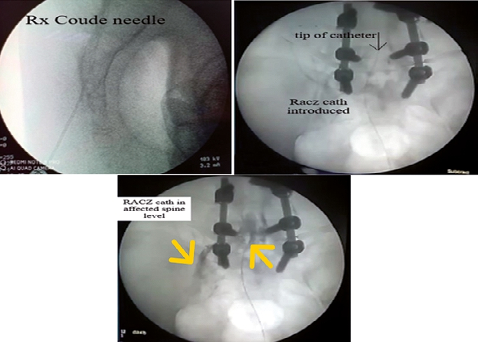
Figure 1. Racz adhesiolysis.
Yellow arrows denote filling of the affected nerve root and the epidural space. and 12 months. The changes in Functional disability using the Oswestry Disability Index at 1, 3, 6, and 12 months. Estimate Pregabalin consumption at 3, 6, and 12 months and Adverse events and complications were also recorded.
Sample size calculation
Sample size was calculated using G*Power 3.1, based on a previous study (Choi et al., 2016) reporting a mean VAS score of 3.9 at 6 months post-adhesiolysis. To detect a difference of 1 point in NRS with SD = 1.1, α = 0.05, and 90% power, at least 25 patients per group were required. We included 30 per group to account for potential dropouts.
Statistical analysis
Data were analyzed using SPSS version
22. Normality was tested with the Shapiro-Wilk test. One-way ANOVA assessed changes over time within groups. Two-way repeated measures ANOVA evaluated between-group differences across timepoints, with Greenhouse-Geisser correction applied when necessary. Post hoc pairwise comparisons were performed using Bonferroni adjustment. Percent improvement in each outcome was calculated as: for NRS and ODI at 6,9 and 12 months. A p-value < 0.05 was considered statistically significant. Improvement = (baseline pre intervention measure-post intervention measure) x 100 (baseline preintervention measure) Figure 2. NAVI adhesiolysis. Yellow arrow denotes filling of the affected nerve root.
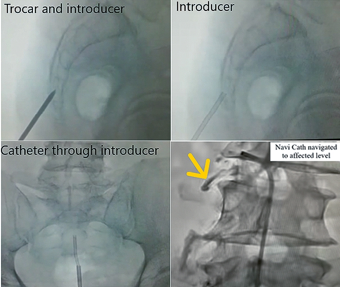
Figure 2. NAVI adhesiolysis. Yellow arrow denotes filling of the affected nerve root.
Patient characteristics
Of the 87 patients enrolled to the study (Figure 3), 60 completed the study (Racz group: 30; Navi-Cath group: 30). Baseline demographic and clinical characteristics were comparable between groups (Table 1).
Table 1. Demographic and clinical data among the studied groups
| Rac’s | Navi | P- value | ||
|---|---|---|---|---|
| Age (years) | 49 ± 6 | 48 ± 7 | 0.7 | |
| Sex | Male | 13 | 14 | 0.5 |
| Female | 17 | 16 | ||
| BMI | 29 ± 4 | 30 ± 5 | 0.6 | |
| Number of previous spine | Once | 19/30 | 18/30 | |
| surgeries | Twice | 10/30 | 10/30 | 0.8 |
| Triple | 1/30 | 2/30 | ||
| spondylosis | 5/30 | 4/30 | 0.5 | |
| DM | 5/30 | 4/30 | 0.6 | |
| Concurrent disease | HTN | 6/30 | 6/30 | 0.5 |
| Mild | 17/30 | 10/30 | ||
| Moderate | 10/30 | 9/30 | ||
| Severe | 2/30 | 9/30 | ||
Mann-Whitney test for non parametric data; t test for parametric data and Chi Square for categoral data.
Pain intensity (NRS)
At baseline, NRS scores did not differ significantly between groups (p = 0.48). No significant differences were observed at 1 month (p = 0.1) or 3 months (p = 0.3). However, the Navi-Cath group showed significantly lower NRS scores at 6 months (p = 0.01) and 12 months (p = 0.002) compared with the Racz group. Both groups demonstrated significant within-group reductions over time (p < 0.001 for each). Pain reduction percentages at 6 and 12 months were significantly greater in the Navi-Cath group (Table 2, Figure 4).
Table 2. NRS ratings across the study groups in relation to time
| NRS | Pre-intervention | 1 M | 3 M | 6 M | 12 M | One way ANOVA | Two-way ANOVA Time*group interaction |
|---|---|---|---|---|---|---|---|
| Racz | 8.9 ± 1 | 2.2 ± 1.2 | 2.5 ± 1.4 | 2.8 ± 1.3 | 3.2 ± 1 | < 0.001 | P = 0.05 |
| NaviCath | 9.1 ± 0.8 0.48 | 1.5 ±1.1 | 2.2 ± 1.1 | 2 ± 0.8 0.01 | 2.4 ± 0.8 0.002 | < 0.001 |
Repeated measure analysis with Bonferroni post hoc analysis; One-way ANOVA is used to compare measures in the same group; Two-way ANOVA for measures comparison between both groups.
Functional disability (ODI)
ODI scores decreased significantly over time in both groups (p < 0.001). The Navi-Cath group consistently demonstrated superior functional recovery compared with the Racz group,
Racz n = 35
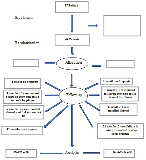
Figure 3. Flow chart. with significant between-group differences at 3, 6, and 12 months (p < 0.001) (Table 4, Figure 5).
Pregabalin consumption
Pregabalin use decreased in both groups compared to baseline. Although consumption was lower in the Navi-Cath group throughout follow-up, the difference was not statistically significant (p = 0.3) (Table 5).
Complications
Adverse events were generally mild and comparable between groups. Reported complications included transient headache (n = 3), temporary motor weakness (n = 2), catheter-related mechanical issues (n = 3 in Racz group only), and coccygodynia (n = 1 with NAVI-Cath). No severe complications (e.g., dural puncture, infection, or hematoma) occurred .No statistical difference was recorded regarding the complication rate between both groups (Table 6).
Discussion
Post-laminectomy syndrome, or failed back surgery syndrome (FBSS), affects up to 40% of patients following lumbar surgery and represents a major cause of chronic pain and disability. Epidural fibrosis is the most frequent underlying mechanism, restricting nerve mobility, impairing movement (Racz et al., 2016). A tethered nerve root is liable to tension, compression, and lack of nutrition Second is postoperative impaired neural circulation, causing ischemic pain (Kim et al., 2013).) Third is venous congestion, which results either from root compression causing outflow obstruction or secondary congestion due to arteriovenous anastomoses or neuroinflammation (Racz et al., 2016 and Kim et al., 2015). However, one other mechanism that could cause FBSS without fibrosis is the presence of the peridural membrane, which is innervated and can become inflamed, particularly in the infra-radicular space (Racz et al., 2016 and Beattie et al., 2000). Two levels of epidural fibrosis were identified by epiduroscpe: dense, resistant fibrous material that was difficult or impossible to penetrate and non-resistant loose or continuous threads and sheets of fibrous material (Bosscher and Heavner 2010). Fibrosis level and the associated vascular changes are correlated to adhesiolysis outcomes (Bosscher and Heavner 2014). These differences could be attributed to the larger size and the steerable mechanism in Navi-Cath, which makes it more effective in dissecting adhesions and fibrosis. Percutaneous adhesiolysis (PA) has proved effective for the treatment of intractable chronic pain after the failure of conservative management and caudal epidural steroid injections (Kim et al., 2021). However, Patients with spinal stenosis or failed back surgery syndrome have a worse response to epidural steroid injection than do herniated disc patients; this is neural blood flow, and limiting the spread of epidurally injected medications. These factors make FBSS particularly resistant to conventional therapies, including pharmacological treatment and epidural steroid injections (Manchikanti et al., 2004). There are several mechanisms by which epidural fibrosis can precipitate pain. One is nerve root tethering, which prevents free mobility of the nerve root in the intra-foraminal and epidural spaces with body because Spinal stenosis and post-spine surgery syndrome are usually associated with irreversible anatomical changes such as epidural fibrosis, scarring, and hypertrophied lateral recess and ligament, which might render the nerve root unresponsive to local steroid application (park et al., 2011; Oh et al., 2019) (Table 3).
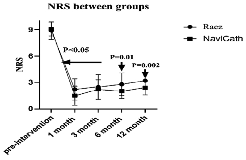
Figure 4. NRS comparison between groups.
Table 3. The oswestry disability index (ODI) between the study groups
| ODI | Pre-intervention | 1 M | 3 M | 6 M | 12 M | One way ANOVA | Two-way ANOVA Time * Group interaction |
|---|---|---|---|---|---|---|---|
| Racz | 45 ± 7 | 36 ± 5 | 32 ± 4 | 29 ± 4 | 30 ± 5 | < 0.001 | P < 0.001 |
| Navi-Cath | 47 ± 5 | 32 ± 4 | 25 ± 5 | 23 ± 4 | 22 ± 3 | < 0.001 | |
| Pos -hoc | 0.1 | 0.01 | < 0.001 | < 0.001 | < 0.001 |
Repeated measure analysis with Bonferroni post hoc analysis; One-way ANOVA for comparison between measures in the same group; Twoway ANOVA for measures comparison between both groups
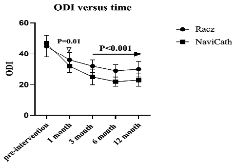
Figure 5. ODI comparison between groups.
Treatment complication Headache Temporary motor weakness Catheter related complication Bending Blocking Steering Coccydynia Mann-Whitney test.
Table 4. Pregabalin consumption between groups
| Pregabalin | Pre-intervention | 3 M | 6 M | 12 M | One way ANOVA | Two-way ANOVA Time*Group interaction |
|---|---|---|---|---|---|---|
| Racz | 300 | 60 ± 16 | 70 ± 28 | 74 ± 22 | > 0.001 | P = 0.3 |
| Navi | 300 | 56 ± 13 | 60 ± 30 | 63±27 | > 0.001 | |
| Post hoc | 0.2 | 0.15 | 0.1 |
Repeated measure analysis with Bonferroni post hoc analysis; One-way ANOVA for comparison between measures in the same group; Twoway ANOVA for measures comparison between both groups.
Table 5. Percent of improvement
| RACZ | Navicath | P Value | |
|---|---|---|---|
| 6 months pain reduction | 68% | 85% | < 0.001 |
| 12 months pain reduction | 63% | 86% | < 0.001 |
| 6 months ODI improvement | 34% | 36% | 0.002 |
| 12 months ODI improvement | 5/% | 54% | < 0.001 |
Chi-square test.
Table 6. Procedure complications
| RACZ | Navi | P- value | ||
|---|---|---|---|---|
| Treatment complication | Headache | 1 | 2 | 0.4 |
| Temporary motor weakness | 1 | 1 | 1 | |
| Catheter related complication | Bending | 2 | 0 | |
| Blocking | 1 | 0 | 0.5 | |
| Steering | 1 | 0 | ||
| Coccydynia | 0 | 1 |
Mann-Whitney test.
To our knowledge, this trial is the first randomized controlled study directly comparing the Racz and Navi-Cath catheters in FBSS patients. Our results demonstrate that both approaches are effective, but Navi-Cath provided more durable pain relief and superior functional recovery at 6 and 12 months. This is consistent with findings by Lee & Lee (2012) that use of a steerable NaviCath catheter improves outcomes in patients refractory to transforaminal epidural injections. The high prevalence of epidural fibrosis in previous reports (Lewik et al., 2023; Yeo et al., 2024) underscores the clinical challenge FBSS poses, especially when fibrotic tissue is severe. These reports provide context for our findings that more precise, steerable instrumentation might overcome some limitations posed by extensive adhesions. Additionally, Quan et al. (2021) demonstrated that outcomes deteriorate with repeated surgical interventions, likely due to worsening fibrosis and adhesions, reinforcing the need for non-surgical interventions that more effectively address epidural fibrosis early. Our study’s results with Navi-Cath suggest that better catheter design might offer longer-term relief with fewer repeat procedures.
Strengths of the study
To the best of current knowledge, this is the first randomized controlled trial directly comparing Racz versus Navi-Cath catheters in FBSS. This addresses a real clinical gap, as prior studies focused on single-catheter outcomes rather than head-to-head comparisons. Prospective, randomized, and double-blind design enhances internal validity.
Clinical implications
These findings suggest that while both Racz and Navi-Cath adhesiolysis improve pain and disability in FBSS, the Navi-Cath catheter may represent the preferred technique for long-term management, particularly in patients with extensive epidural fibrosis or difficult-to-access lesions. Importantly, complication rates did not differ between groups, supporting the safety of both techniques.
Study limitations
This trial has several limitations. It was conducted in a single center with a modest sample size, which may limit external validity. Follow-up was limited to 12 months, precluding long-term outcome assessment. The absence of a conservative control group (e.g., epidural steroid injections alone) restricts broader comparative analysis. Pregabalin use was the only measure of pharmacologic need, and additional analgesic metrics or quality-of-life assessments would strengthen the evaluation. Finally, patients were not stratified by fibrosis severity, which could influence treatment response.
Recommendations
Future multicenter randomized trials with larger cohorts and longer follow-up are needed to validate these findings. Incorporating imaging-based fibrosis grading or epiduroscopic classification may help tailor catheter selection. Cost-effective-
ness analyses are warranted to guide resource allocation. Finally, integrating patient-reported outcomes and psychological assessments would provide a more comprehensive view of clinical benefit.
Conclusion
In this randomized clinical trial, both Racz and Navi-Cath catheters improved pain and disability in FBSS patients. However, Navi-Cath produced significantly superior outcomes at 6 and 12 months, with sustained pain relief, functional recovery, and no increase in complication rates. These findings support the use of steerable catheters as a potentially more effective strategy for percutaneous adhesiolysis in FBSS.
Referencias
1. Lewik G, Glinkowski W, Krawczak K, Serzysko B, Rola R. Postoperative Epidural Fibrosis: Challenges and Opportunities A Review. J Pain Res. 2023;16:2967-2981. https://doi.org/10.22603/ssrr.20230106 PMID: https://pubmed.ncbi.nlm.nih.gov/38618214.
2. Quan M, Song J, Yang Y, Ma Y, Zhang S, Sun Y. Analysis of pain markers and epidural fibrosis caused by revision surgery of lumbar vertebrae. BMC Musculoskelet Disord. 2021;22(1):
28. https://doi.org/10.1186/s12891-020-03920-z PMID: https://pubmed.ncbi.nlm.nih.gov/33402133.
3. Yeo J, Lee HS, Kim Y, Kim YS, Kim DH, Kim YH, et al. Failed back surgery syndrome-terminology, etiology, prevention, evaluation, and management: a narrative review. Pain Med. 2024;25(4):610620. https://doi.org/10.12701/jyms.2024.00339 PMID: https://pubmed.ncbi.nlm.nih.gov/38853538.
5. Lee JH, Lee SH. Clinical effectiveness of percutaneous adhesiolysis using NaviCath for the management of chronic pain due to lumbosacral disc herniation. Pain Physician. 2012;15(3):213-221. https://doi.org/10.36076/ppj.2012/15/213 PMID: https://pubmed.ncbi.nlm.nih.gov/22622905.
6. Brito-Garcia, N., Garcia-Perez, L., Kovacs, F. M., del Pino-Sedeno, T., Perez-Ramos, J., Imaz-Iglesia, I., & Serrano-Aguilar, P. Efficacy, effectiveness, safety, and cost-effectiveness of epidural adhesiolysis for treating failed back surgery syndrome. A systematic review. Pain Medicine2019; 20(4), 692-706.
7. Hossieni, B., Dadkhah, P., Moradi, S., Hashemi, S. M., & Safdari, F. The results of treating failed back surgery syndrome by adhesiolysis: comparing the one-and three-day protocols. Anesthesiology and Pain Medicine, 2017, 7.5. https://doi.org/10.5812/aapm.60271 PMID: https://pubmed.ncbi.nlm.nih.gov/29696119.
8. Thomson, S. Failed back surgery syndrome-Definition, epidemiology and demographics. BJP 2013, 7, 56-59. https://doi.org/10.1177/2049463713479096. PMID: https://pubmed.ncbi.nlm.nih.gov/26516498.
9. Rigoard, P.; Desai, M.; Taylor, R.S. Failed back surgery syndrome: What’s in a name? A proposal to replace “FBSS” by “POPS” Neurochirurgie 2015, 61, S16-S21.
10. Kim, J. Y., Lee, Y. H., Yoo, S., Kim, J. Y., Joo, M., & Park, H. J. Factors predicting the success of adhesiolysis using a steerable catheter in lumbar failed back surgery syndrome: a retrospective study. Journal of Clinical Medicine.2021; 10(5), 913. https://doi.org/10.3390/jcm10050913 PMID: https://pubmed.ncbi.nlm.nih.gov/33652702.
11. Choi, S. S., Lee, J. H., Kim, D., Kim, H. K., Lee, S., Song, K. J. & Shim, J. H. Effectiveness and factors associated with epidural decompression and adhesiolysis using a balloon-inflatable catheter in chronic lumbar spinal stenosis: 1-year follow-up. Pain medicine.2016; 17(3), 476-487. https://doi.org/10.1093/pm/pnv018 PMID: https://pubmed.ncbi.nlm.nih.gov/26814254.
12. Manchikanti L, Rivera JJ, Pampati V, Damron KS, McManus CD, Brandon DE, Wilson SR. One day lumbar epidural adhesiolysis and hypertonic saline neurolysis in treatment of chronic low back pain: a randomized, double-blind trial. Pain Physician. 2004 Apr 1;7(2):177-86. https://doi.org/10.36076/ppj.2004/7/177 PMID: https://pubmed.ncbi.nlm.nih.gov/16868590.
13. Racz SH, Gerdesmeyer L, Justiz R, Hayek SM, Kaplan ED, El Terany MA, Knezevic NN. Percutaneous and endoscopic adhesiolysis in managing low back and lower extremity pain: a systematic review and meta-analysis. Pain Physician. 2016;19(2):E245. https://doi.org/10.36076/ppj/2016.19.e245
14. Kim SH, Choi WJ, Suh JH, Jeon SR, Hwang CJ, Koh WU, Lee C, Leem JG, Lee SC, Shin JW. Effects of transforaminal balloon treatment in patients with lumbar foraminal stenosis: A randomized, controlled, double-blind trial. Pain Physician. 2013; 16:213-224. https://doi.org/10.36076/ppj.2013/16/213 PMID: https://pubmed.ncbi.nlm.nih.gov/23703408.
15. Kim J, Jund HJ, Nahn FS, Lee PB. Does improvement in epidurography following percutaneous epidural neuroplasty correspond to patient outcome? Pain Practice. 2015; 15:407-413. https://doi.org/10.1111/papr.12197 PMID: https://pubmed.ncbi.nlm.nih.gov/24750546.
16. Beattie PF, Meyers SP, Stratford P, Millard RW, Hollenberg GM. Associations between patient report of symptoms and anatomic impairment visible on lumbar magnetic resonance imaging. Spine. 2000; 25:819-828. https://doi.org/10.1097/00007632-20000401000010 PMID: https://pubmed.ncbi.nlm.nih.gov/10751293.
17. Bosscher HA, Heavner JE. Incidence and severity of epidural fibrosis after back surgery: An endoscopic study. Pain Practice. 2010; 10:18-24. https://doi.org/10.1111/j.1533-2500.2009.00311.x. PMID: https://pubmed.ncbi.nlm.nih.gov/19735365.
18. Bosscher H, Heavner JE. Lumbosacral epiduroscopy findings predict treatment outcome. Pain Practice. 2014; 14:506-5014. https://doi.org/10.1111/papr.12112 PMID: https://pubmed.ncbi.nlm.nih.gov/24118805.
19. Oh, Y.; Kim, D.-H.; Park, J.-Y.; Ji, G.Y.; Shin, D.A.; Lee, S.W.; Park, J.K.; Shin, J.-W.; Choi, S.-S. Factors Associated with Successful Response to Balloon Decompressive Adhesiolysis Neuroplasty in Patients with Chronic Lumbar Foraminal Stenosis. J. Clin. Med. 2019, 8, 1766. https://doi.org/10.3390/jcm8111766 PMID: https://pubmed.ncbi.nlm.nih.gov/31652838.
20. Park, C.H.; Lee, S.H.; Jung, J.Y. Dural sac cross-sectional area does not correlate with efficacy of percutaneous adhesiolysis in single level lumbar spinal stenosis. Pain Physician 2011, 14, 377382. https://doi.org/10.36076/ppj.2011/14/377 PMID: https://pubmed.ncbi.nlm.nih.gov/21785481.