
Enrique Sepúlveda Haro 1 ,* , Salvador Romero Molina 1 , Luis Morcillo Hidalgo 2 , Rafael Méndez Natera 2 , Arancha Díaz Expósito 2 , Antonia Pilar Martín de la Rosa 2
 ORCID
ORCIDRecibido: 13-10-2021
Aceptado: 12-11-2021
©2022 El(los) Autor(es) – Esta publicación es Órgano oficial de la Sociedad de Anestesiología de Chile
Revista Chilena de Anestesia Vol. 51 Núm. 3 pp. 261-262|https://doi.org/10.25237/revchilanestv5115031055
PDF|ePub|RIS
Dear Editor,
The insertion of a transesophageal echocardiography (TEE) probe is usually performed in a blind manner both in the anesthetized and awake patient, but it is often hampered by a lack of tolerance of the patient or by oropharyngoesophageal anatomical abnormalities. Indirect videolaryngoscopy is useful in the guidance of TEE probe insertion in the anesthetized intubated patient[1],[2], but it has not been used in the sedated, non intubated patient to date. Of note, in the awake sedated patient with difficult airway, videolaryngoscopy has shown to guide intubation with success[3].
We present here the clinical case of a difficult TEE probe insertion that was successfully achieved with the guidance of an Airtraq® videolaryngoscope (AV) (Figure 1) in a patient under sedation.
The patient was a male, 87 years old, 65 kg, ASA grade 4, who was scheduled for transesophageal echocardiography as an outpatient. He had hypertension, severe chronic kidney disease and cardiac insufficiency with preserved ejection fraction secondary to severe mixed aortic valve disease, ischemic cardiomyopathy and atrial fibrillation. He had a multifactorial anaemia, presented a Mallampati class 3 and was edentulous. The study aimed to evaluate the anatomical situation of the left atrial appendage, as part of the procedure planning of the endovascular closure intervention he was going to be scheduled.
Standard monitorization, an intravenous catheter, oxygen supplementation through nasal cannula and a bite block were placed. Light sedation was achieved with intravenous midazolam 2 mg and topicalization of the oral cavity with sprayed lidocaine 10% was applied. Insertion of the TEE probe was tried several times by an experienced cardiologist with good patient
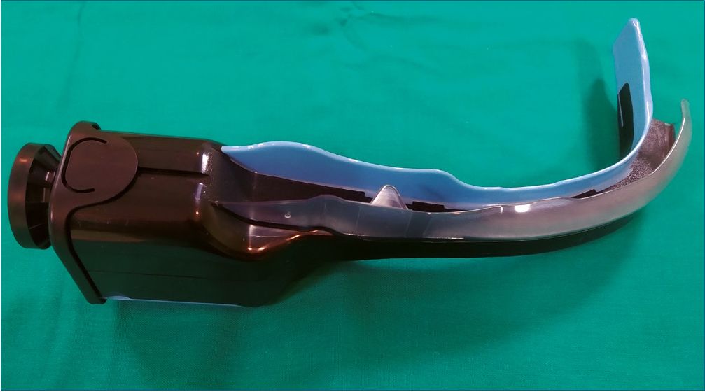
Figure 1. Airtraq® videolaryngoscope.
collaboration, but it could not be placed in the esophagus, as an excessive resistance was felt at 15 cm depth from the lips with every attempt.
In this setting, sedation was increased with additional intravenous midazolam 7 mg, obtaining a deep level of sedation, but TEE probe introduction was neither achieved. An anesthetist was asked for assessment, who found no relevant airway alteration, with a normal mouth opening, a relatively limited neck extension and normal thyromental and thyrohyoid distances. The patient was placed supine and a after manually opening the mouth of the patient, insertion of both the regular size AV (size 3) and TEE probe was achieved easily in this context of sedation and complete absence of teeth. Laryngoscopy with AV was very well tolerated, obtaining a pharyngolaryngeal view as good as the one in anesthetized, intubated patients, and normal glottic structures without any evident pharyngeal alteration could be seen. The TEE probe was then advanced on the right side of the AV (outside the tube-guiding channel) until it could be seen from the AV view. The tip of the TEE probe was passed aiming the left lateral and posterior area of the laryngopharynx, where no obstruction was felt and full insertion of the TEE probe could be performed with good patient tolerance.
The patient was placed again in the left lateral decubitus position and the TEE study was performed with hemodynamic and respiratory stability, patient comfort was acceptable for the procedure and he was discharged from hospital a few hours later.
Anesthesia support may be needed in some TEE procedures when patients do not cooperate, deep sedation is required or hemodynamic or respiratory instability is anticipated. Ambulatory TEE is routinely performed in our center under conscious sedation using midazolam as a sole drug by the Cardiology Team. In this particular case, the patient received a high dose of intravenous midazolam (9 mg) that could potentially have caused respiratory depression. After achieving a deep sedation and with the unability to introduce the TEE probe, an anatomical abnormality was suspected and Anesthesiology support was asked. The anesthesiologist assessment found no apparent anatomical abnormality and an indirect laryngoscopic exploration under that situation of deep sedation was decided.
Hirabayashi Y et al. presented in 2008 a method for Airtraq®– assisted TEE probe insertion in an anesthetized and intubated patient[1]. Recently, a systematic review and meta-analysis of randomized controlled trials has shown that videolaryngoscopy during TEE probe insertion in the anesthetized and intubated patient is safe compared with other methods such as blind insertion and direct laryngoscopy guidance[2]. As a technical detail, in our case, we inserted the TEE probe through the mouth of the patient but outside the tube-guiding channel in order to allow a higher range of motion, rather than through the channel, as Hirabayashi Y et al did, and this could have been easy in our case because of the lack of teeth of our patient[1].
It is evident that the deep degree of sedation observed in our patient helped with tolerance for both Airtraq® videolaryngoscope and TEE probe insertion, but we speculate that patients with lighter level of sedation could potentially tolerate this procedure, as it has been observed in similar situations regarding videolaryngoscope-guided intubation in awake patients under moderate sedation[3].
To our knowledge, this is the first reported case of a videolaryngoscopeguided TEE probe insertion in a sedated, non-intubated patient. We speculate that Airtraq® videolaryngosco- pe guidance may be an alternative when difficult insertion of TEE probe is found even in the awake patient under moderate degrees of sedation, although further studies are needed to evaluate the safety and effectivity of this technique.
Acknowledgements: We would like to thank Dr. Mínguez Mañanes, Anesthesiologist, Department of Anesthesiology and Critical Care, Virgen de la Victoria University Hospital, Málaga, Spain, for teaching us Airtraq® videolaryngoscope management.
References
1. Hirabayashi Y, Okada O, Seo N. Airtraq laryngoscope for the insertion of a transesophageal echocardiography probe. J Cardiothorac Vasc Anesth. 2008 Apr;22(2):331–2. https://doi.org/10.1053/j.jvca.2007.06.012 PMID:18375345
2. Namekawa M, Tsujimoto Y, Banno M, Kataoka Y, Tsujimoto H, Inaba Y, et al. Videolaryngoscopy for transesophageal echocardiography probe insertion: a systematic review and meta-analysis of randomized controlled trials. J Anesth. 2020 Jun;34(3):453–63. https://doi.org/10.1007/s00540-020-02759-x PMID:32219541
3. Tai F, Ho Y, Cheng B, Mui K, Chan M, Wong O. The use of the Airtraq® in awake tracheal intubation: a case series. Hong Kong J Emerg Med. 2015;22(4):248–55. https://doi.org/10.1177/102490791502200407.