Ricardo Serna Muñoz1, Nora Bernal Ríos1, María García Bravo1
 ORCID
ORCID
Recibido:
Aceptado: 2026-01-15
©2026 El(los) Autor(es) – Esta publicación es Órgano oficial de la Sociedad de Anestesiología de Chile
Revista Chilena de Anestesia Vol. 39 Núm. 5 | https://doi.org/10.25237/revchilanestv55n5-26
PDF
Intubación nasofibroscópica despierto en un paciente de alto riesgo con dificultades anatómicas y fisiológicas combinadas: Posradioterapia, trismus, EPOC y estenosis carotídea severa
Abstract
Airway management in patients with prior head and neck malignancies, radiation-induced anatomical changes, and significant comorbidities poses major challenges. We present the case of a 76-year-old woman with a history of oral squamous cell carcinoma treated with mandibulectomy and radiotherapy, who developed severe trismus and was admitted with otomastoiditis requiring urgent surgical drainage. Her medical background included GOLD E chronic obstructive pulmonary disease, severe carotid artery stenosis, epilepsy, and chronic oncologic pain. Imaging confirmed advanced sinus and mastoid involvement. Due to the patient’s limited mouth opening (< 1 cm), altered anatomy, and high perioperative risk, she was classified as a physiologically and anatomically difficult airway. A plan for awake nasal fiberoptic intubation was executed using a dexmedetomidine-fentanyl sedation protocol combined with stepwise topical anesthesia. Multimodal monitoring included cerebral oximetry and invasive arterial pressure monitoring, allowing hemodynamic support with norepinephrine. After successfully securing the airway, general anesthesia was induced, and the surgical procedure proceeded uneventfully. The patient was extubated fully awake, transferred to the intermediate care unit for postoperative monitoring, and discharged without complications. This case underscores the importance of individualized planning based on current international guidelines for difficult airway management, particularly in high-risk patients with trismus and prior radiotherapy. Awake nasal fiberoptic intubation remains the safest and most effective strategy in such scenarios. When performed by experienced personnel within a structured perioperative protocol, it allows safe airway control while minimizing complications. Predefined rescue strategies and close interdisciplinary collaboration are key components in achieving favorable outcomes in complex airway cases.
Resumen
Airway management in patients with prior head and neck malignancies, radiation-induced anatomical changes, and significant comorbidities poses major challenges. We present the case of a 76-year-old woman with a history of oral squamous cell carcinoma treated with mandibulectomy and radiotherapy, who developed severe trismus and was admitted with otomastoiditis requiring urgent surgical drainage. Her medical background included GOLD E chronic obstructive pulmonary disease, severe carotid artery stenosis, epilepsy, and chronic oncologic pain. Imaging confirmed advanced sinus and mastoid involvement. Due to the patient’s limited mouth opening (< 1 cm), altered anatomy, and high perioperative risk, she was classified as a physiologically and anatomically difficult airway. A plan for awake nasal fiberoptic intubation was executed using a dexmedetomidine-fentanyl sedation protocol combined with stepwise topical anesthesia. Multimodal monitoring included cerebral oximetry and invasive arterial pressure monitoring, allowing hemodynamic support with norepinephrine. After successfully securing the airway, general anesthesia was induced, and the surgical procedure proceeded uneventfully. The patient was extubated fully awake, transferred to the intermediate care unit for postoperative monitoring, and discharged without complications. This case underscores the importance of individualized planning based on current international guidelines for difficult airway management, particularly in high-risk patients with trismus and prior radiotherapy. Awake nasal fiberoptic intubation remains the safest and most effective strategy in such scenarios. When performed by experienced personnel within a structured perioperative protocol, it allows safe airway control while minimizing complications. Predefined rescue strategies and close interdisciplinary collaboration are key components in achieving favorable outcomes in complex airway cases.
Introduction
Airway management in patients with prior head and neck cancer surgery poses significant challenges for anesthesiologists. Anatomical distortion, fibrosis from surgery and radiotherapy, limited mouth opening, and ongoing infections may all contribute to anatomical changes that increase the complexity of the airway management. These cases require detailed preoperative planning, careful sedation strategies, and often the use of awake intubation techniques to maintain spontaneous ventilation and airway reflexes[1],[2].
Patients with advanced comorbidities including chronic obstructive pulmonary disease (COPD), structural epilepsy, cerebrovascular disease, and chronic opioid use present additional physiological complexity[3],[4]. The risk of respiratory depression, hemodynamic instability, and perioperative neurological deterioration must be balanced against the need for surgical airway access and infection control[5].
This case describes the perioperative management of a 76-year-old woman with a history of tongue and mandibular cancer, with mandibular reconstruction, chronic otomastoiditis, and multiple systemic comorbidities. It highlights the use of awake fiberoptic nasal intubation, multimodal neuromonitoring, and individualized hemodynamic control to ensure airway safety and minimize perioperative risk.
Case presentation
A 76-year-old woman was admitted to the emergency department with a 48-hour history of right-sided periauricular and facial pain, foul-smelling purulent otorrhea, and ipsilateral erythema. Her medical history was notable for GOLD E COPD on long-term oxygen therapy, multi-infarct cerebrovascular disease (including left cerebellar infarcts and prior intracerebral hemorrhage), epilepsy secondary to structural lesions, and chronic oncologic pain. She also had a history of oral squamous cell carcinoma: tongue cancer 25 years earlier and mandibular cancer 7 years prior, the latter treated with mandibulectomy, flap reconstruction, 7 cycles of cisplatin-based chemotherapy, and 35 sessions of radiotherapy.
She had recurrent episodes of sinusitis and otomastoiditis. Pseudomonas aeruginosa was isolated from sinus aspirates in march 2025, prompting prolonged outpatient treatment with oral ciprofloxacin, which failed to achieve complete resolution. Given the clinical deterioration and persistent purulent otorrhea, she was admitted for intravenous antibiotic therapy with meropenem and linezolid, along with oral vancomycin prophylaxis due to a history of Clostridioides difficile colitis. An urgent right inferior turbinoplasty, middle ear debridement, and placement of a tympanostomy tube for drainage and culture collection was scheduled by otolaryngology.
On physical examination, the patient was awake, alert, and fully oriented, with intact higher mental functions and no signs of acute neurological deficit. Cranial nerve assessment revealed right-sided central facial nerve paresis, consistent with prior cerebrovascular events. Notably, mouth opening was severely restricted to less than 1 cm, likely due to post-radiation fibrosis and prior mandibular reconstruction, representing a significant concern for airway access. Purulent otorrhea was observed from the right external auditory canal. Pulmonary examination revealed decreased breath sounds bilaterally, consistent with underlying emphysematous changes. Cardiovascular and abdominal examinations were unremarkable. Muscle strength was preserved in all four extremities, with symmetric tone and no focal motor deficits.
A CT scan of paranasal sinuses revealed opacification of the right maxillary, ethmoidal, and sphenoidal sinuses, with erosion of the maxillary sinus floor and possible oroantral communication. Hypodensities were also observed in the right middle ear and mastoid cells (Figure 1).
Doppler ultrasound revealed significant stenosis of the right common carotid artery due to irregular atheroma, without hemodynamic compromise (Figure 2).
Given this combination of anatomical, infectious, vascular, neurologic, and pharmacologic risk factors, the patient was classified as high risk for airway and perioperative cardiovascular, neurologic and respiratory complications.
Following a thorough medical evaluation, the surgical and anesthesia teams discussed the airway management plan and agreed on an awake nasal fiberoptic intubation. The procedure was clearly explained to the patient and her family, and informed consent was obtained. She was transferred to the operating room under supplemental oxygen via nasal cannula at 5 L/min, fully oriented and neurologically intact. Standard monitoring was initiated, including non-invasive blood pressure, electrocardiography, pulse oximetry, plethysmography, bilateral cerebral oximetry, and a SedLine EEG monitor. In addition, a right radial arterial line was placed for continuous blood pressure monitoring.
Topical airway preparation included nebulized 2% lidocaine and vasoconstrictor application to the right nostril. Sedation was initiated with dexmedetomidine at 2 mcg/kg/h (total dose: 25 mcg) and fentanyl at 1 mcg/kg. The nasoand oropharynx were further anesthetized with 10% lidocaine spray. Using the “spray-as-you-go” technique, lidocaine was administered thru the nasofibroscope working channel. With the patient in a seated position the scope was introduced through the left nostril, advanced through the upper airway, guided past the vocal cords and advanced until the carina was visualized, allowing for the successful placement of a 6.0 mm internal diameter nasotracheal tube. Correct positioning was confirmed by capnography and auscultation.
General anesthesia was then induced with 20 mg of propofol, additional 100 mcg of fentanyl, and 40 mg of rocuronium, with a norepinephrine infusion initiated at 0.05 mcg/kg/min and titrated to maintain mean arterial pressure within 20% of baseline and cerebral oximetry variations below 10% of initial saturation values. Surgical drainage proceeded without complications; estimated blood loss was 30 mL. Upon completion, the patient was extubated fully awake and transferred to the intermediate care unit for continued neurological, cardiovascular, and respiratory monitoring.
In the following days, postoperative pain was managed with an oxycodone IV home-pump infuser delivering 6 mg over 24 hours, which was discontinued after completing the planned dose. The patient showed favorable clinical evolution, remained symptoms free, and was discharged home without complications.
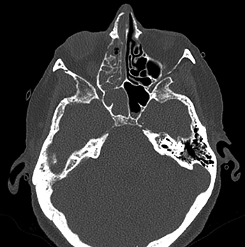
Figure 1
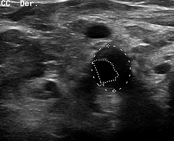
Figure 2.
Discussion
Airway management in high-risk patients requires careful consideration of both anatomical and physiological challenges[3]. In this case, the decision to proceed with awake tracheal intubation is consistent with current international guidelines recommendations. The 2022 American Society of Anesthesiologists (ASA) practice guidelines and the 2020 Difficult Airway Society (DAS) consensus on awake tracheal intubation recommend an awake approach when difficult ventilation, high aspiration risk, or challenging invasive rescue access are anticipated[5]-[7]. In our case, the choice of awake fiberoptic nasal intubation, while maintaining spontaneous ventilation, using stepwise topical anesthesia, and ensuring immediate availability of rescue strategies, reflects the importance of protocolized airway management in high-risk patients.
In head and neck cancer patients with severe trismus and post-radiotherapy fibrosis, airway management is particularly challenging due to profound anatomical restrictions. With an interincisor opening of less than 1 cm, the insertion of a conventional laryngoscope or even supraglottic airway devices become technically unfeasible, and oral manipulation with a fiberscope is extremely limited[8],[9].
In such scenarios, the nasal route provides the most reliable access, allowing the passage of the endoscope while maintaining spontaneous ventilation and patient cooperation, as well as preserving the surgical field[10]-[12]. Although epistaxis, abundant secretions and nasal trauma are recognized risks of this approach, their incidence can be minimized through careful preparation, including topical vasoconstrictors, nebulized lidocaine for mucosal anesthesia, and the selection of a small-caliber endotracheal tube (6.0). These measures in our case contributed to a smooth intubation procedure without bleeding, reinforcing that the nasal awake fiberoptic technique represents the safest and most effective strategy in patients with such severe anatomical limitations.
Beyond the anatomical considerations, this case also met criteria for a physiologically difficult airway. Severe COPD requiring continuous home oxygen conferred a high risk of rapid desaturation with even brief apneic periods, and chronic lung disease is frequently accompanied by secondary pulmonary hypertension and right ventricular dysfunction, increasing the likelihood of hemodynamic instability during airway instrumentation[3],[13]-[15]. These comorbidities mandate an awake strategy that preserves spontaneous ventilation and minimized the probability of hypoxemia or physiologic instability. Concomitantly, the history of cerebrovascular disease and severe carotid stenosis underscored the need to safeguard cerebral and myocardial perfusion: even short hypotensive episodes during sedation or induction could compromise cerebral blood flow and/or myocardial oxygen delivery. Accordingly, we inserted an arterial catheter and employed continuous cerebral oximetry to guide precise titration of anesthetic agents; a norepinephrine infusion was maintained and titrated to ensure stable perfusion pressures, neck positioning was kept neutral to avoid carotid compromise, and induction was deliberately slow and stepwise to avoid abrupt hemodynamic fluctuations[16]-[18]. These monitoring measures aimed to preserve both systemic and cerebral oxygenation in this high-risk patient.
In anticipation that post-radiotherapy laryngeal fibrosis can thwart even a well-prepared awake fiberoptic intubation, we predefined a rescue pathway consistent with international guidance. Within the DAS framework, an unsuccessful attempt at awake tracheal intubation (ATI) is the unplanned removal of the flexible bronchoscope, videolaryngoscope, or tracheal tube from the airway, and failed ATI is the inability to achieve tracheal intubation after three attempts by the primary operator plus one additional attempt by a more experienced operator (3 + 1) [5].
Our staged plan prioritized oxygenation and minimization of airway trauma: (i) immediate call for help and re-optimization (re-topicalization, operator change), (ii) switch to the contralateral nostril, downsize the tube (e.g., 5.5-6.0), and maintain oxygenation with high-flow nasal oxygen while preserving spontaneous ventilation; given the < 1 cm interincisor distance, oral videolaryngoscopy/supraglottic rescue were not viable. If ATI met failed criteria (3 + 1), we would avoid ablation of spontaneous ventilation and proceed to an elective awake front-of-neck airway (tracheostomy/cricothyrotomy) with ENT support; an asleep strategy was considered only as a last resort if oxygenation could be reliably secured. This pre-committed sequence aligns with the DAS “unsuccessful ATI” algorithm and ASA principles that place oxygenation and attempt limits at the center of decision-making[5],[7],[19],[20].
This case translates guideline principles into operational steps that directly mitigate hypoxemia risk and hemodynamic instability in a high-risk patient. This approach aligns and operationalizes airway guideline recommendations into reproducible actions that are auditable in real time and scalable across teams.
Guidelines from bodies such as the ASA (2022) and DAS (2025) are a pillar of safe airway management, but their impact depends on thoughtful individualization to the patient and context. Translating recommendations to practice requires weighing anatomic feasibility, physiologic reserve, comorbidities, team expertise, and resource constraints. This balance standardizing and individualizing is what converts high-level guidance into bedside safety for high-risk patients[5],[7],[21],[22].
Context also matters: site-of-care and resource availability decisively shape what is feasible and safe. The same plan may differ between a tertiary operating room with ENT standby, flexible bronchoscopes, high-flow nasal oxygen, cerebral oximetry, and ready front-of-neck kits, versus a resource-limited or ambulatory setting without those supports. Pre-procedural appraisal should therefore include team expertise and staffing, equipment redundancy (bronchoscopes, suction, small-caliber nasal tubes, vasoconstrictors), monitoring capability (arterial line, rSO2), rescue pathways (FONA drills, ENT availability), postoperative disposition (ICU step-down access), and logistics (oxygen supply, space for airway cart).[23]-[28]. When critical elements are lacking, the plan should be modified or the venue escalated (e.g., schedule in a higher-acuity OR, secure ENT support, or lower the threshold for an elective awake front-of-neck strategy). Guidelines provide the scaffold; contextualization and resource-aware planning convert that scaffold into safe, reproducible care for high-risk patients.
Ethical approval
This study was approved by the institutional Research Ethics Committee XYZ, in a meeting held on month day year, as recorded in act number #.
Protection of human and animal subjects
The authors declare that no experiments were performed on humans or animals for this study. The authors declare that the procedures followed were in accordance with the regulations of the relevant clinical research ethics commit-tee and with those of the Code of Ethics of the World Medical Association (Declaration of Helsinki).
Confidentiality of data
The authors declare that they have followed the protocols of their work center on the publication of patient data.
Right to privacy and informed consent
The authors declare that no patient data appear in this article. The authors have obtained the written informed consent of the patients or subjects mentioned in the article. The corresponding author is in possession of this document.
Acknowledgements: Authors contributions: RSM Study
planning, data collection, interpretation of resultas and initial writing of the manuscript, medical attention and case management NBR, study planning, Medical attention and case management, interpretation of results and edition of the manuscript, MGB Study planning, edition of the manuscript, medical attention. · Assistance for the study: None. · Financial support and sponsorship: None. · Appreciation: None.
Referencias
1. Sandefur BJ, Driver BE, Long B. Managing Awake Intubation. Annals of Emergency Medicine. Elsevier Inc.; 2024.
2. Huitink JM, Buitelaar DR, Schutte PFE. Awake fibrecapnic intubation: A novel technique for intubation in head and neck cancer patients with a difficult airway. Anaesthesia. 2006 May;61(5):449-52. https://doi.org/10.1111/j.1365-2044.2006.04586.x PMID: https://pubmed.ncbi.nlm.nih.gov/16674619.
3. Jabaley CS. Managing the Physiologically Difficult Airway in Critically Ill Adults. Vol. 27, Critical Care. BioMed Central Ltd; 2023. https://doi.org/10.1186/s13054-023-04371-3 PMID: https://pubmed.ncbi.nlm.nih.gov/36941620.
4. Gostelow N, Yeow D. Awake tracheal intubation: a narrative review. Vol. 2, Journal of Oral and Maxillofacial Anesthesia. AME Publishing Company; 2023. https://doi.org/10.21037/joma-23-17
5. Ahmad I, El-Boghdadly K, Bhagrath R, Hodzovic I, McNarry AF, Mir F, et al. Difficult Airway Society guidelines for awake tracheal intubation (ATI) in adults. Anaesthesia. 2020 Apr 1;75(4):509-28. https://doi.org/10.1111/anae.14904. PMID: https://pubmed.ncbi.nlm.nih.gov/31729018.
6. Rosenblatt WH, Yanez ND. A Decision Tree Approach to Airway Management Pathways in the 2022 Difficult Airway Algorithm of the American Society of Anesthesiologists. Anesth Analg. 2022 May 1;134(5):910-5. https://doi.org/10.1213/ane.0000000000005930 PMID: https://pubmed.ncbi.nlm.nih.gov/35171880.
7. Apfelbaum JL, Hagberg CA, Connis RT, Abdelmalak BB, Agarkar M, Dutton RP, et al. 2022 American Society of Anesthesiologists Practice Guidelines for Management of the Difficult Airway. Anesthesiology. 2022 Jan 1;136(1):31-81. https://doi.org/10.1097/aln.0000000000004002 PMID: https://pubmed.ncbi.nlm.nih.gov/34762729.
8. Akazawa M, Shimizu M, Fujino Y, Kato H. Radiation-Induced Nasopharyngeal Fibrosis Resulting in a Difficult Airway: A Case Report. Cureus. 2025 Feb 17. https://doi.org/10.7759/cureus.79130 PMID: https://pubmed.ncbi.nlm.nih.gov/40109800.
9. Lu WC, Wu ZF, Lai HC. Successful Awake Video Stylet Orotracheal Intubation in an Oral Cancer Patient with Limited Mouth Opening and Radiation Fibrosis. Vol. 87, Indian Journal of Surgery. Springer; 2025. p. 409-10. https://doi.org/10.1007/s12262-024-04090-1
10. Pirlich N, Noppens RR. Local airway anaesthesia for awake fibreoptic intubation. Vol. 10, Trends in Anaesthesia and Critical Care. Churchill Livingstone; 2016. p. 22-8. https://doi.org/10.1016/j.tacc.2016.10.002
11. Badiger S, John M, Fearnley RA, Ahmad I, Asai T. Optimizing oxygenation and intubation conditions during awake fibre-optic intubation using a high-flow nasal oxygen-delivery system. Br J Anaesth. 2015 Oct 1;115(4):629-32. https://doi.org/10.1093/bja/aev262 PMID: https://pubmed.ncbi.nlm.nih.gov/26253608.
12. Hyman JB, Rosenblatt WH. Awake Intubation Techniques, and Why It Is Still an Important Skill to Master. Vol. 12, Current Anesthesiology Reports. Springer; 2022. p. 382-9. https://doi.org/10.1007/s40140-022-00529-x
13. Fonseca D, Graça MI, Salgueirinho C, Pereira H. Physiologically difficult airway: How to approach the difficulty beyond anatomy. Vol. 48, Trends in Anaesthesia and Critical Care. Churchill Livingstone; 2023. https://doi.org/10.1016/j.tacc.2023.101212
14. Mosier JM, Joshi R, Hypes C, Pacheco G, Valenzuela T, Sakles JC. The physiologically difficult airway. Vol. 16, Western Journal of Emergency Medicine. eScholarship; 2015. p. 1109-17. https://doi.org/10.5811/westjem.2015.8.27467 PMID: https://pubmed.ncbi.nlm.nih.gov/26759664.
15. Vakil B, Baliga N, Myatra S. The physiologically difficult airway. Airway. 2021;4(1): 4. https://doi.org/10.4103/arwy.arwy_10_21
16. Hejgl A, Jiránková & K, Malucelli & A, Sejkorová & A, Radovnický & T, Bartoš & R, et al. Selective internal carotid artery cross-clamping increases the specificity of cerebral oximetry for indication of shunting during carotid endarterectomy. Acta Neurochir (Wien) [Internet]. 2020;163:1807-17. Available from:. https://doi.org/10.1007/s00701-020-04621-1 PMID: https://pubmed.ncbi.nlm.nih.gov/33106902.
17. Denault A, Deschamps A, Murkin JM. A proposed algorithm for the intraoperative use of cerebral near-infrared spectroscopy. In: Seminars in Cardiothoracic and Vascular Anesthesia. 2007. p. 274-81. https://doi.org/10.1177/1089253207311685 PMID: https://pubmed.ncbi.nlm.nih.gov/18270192.
18. Murkin JM, Arango M. Near-infrared spectroscopy as an index of brain and tissue oxygenation. Br J Anaesth. 2009;103. https://doi.org/10.1093/bja/aep299 PMID: https://pubmed.ncbi.nlm.nih.gov/20007987.
19. Duggan L V. 4th National Audit Project of The Royal College of Anaesthetists and The Difficult Airway Society (NAP4) Major Complications of Airway Management in the United Kingdom. Canadian Journal of Anesthesia/Journal canadien d’anesthésie. 2011 Nov;58(11):1061-2. https://doi.org/10.1007/s12630-011-9576-5
20. Heidegger T. Management of the Difficult Airway. Longo DL, editor. New England Journal of Medicine [Internet]. 2021 May 13;384(19):1836-47. Available from: http://www.nejm.org/doi/10.1056/NEJMra1916801
21. Evans A, Morton B, Groom P. Difficult Airway Society guidelines for awake tracheal intubation in adults is lidocaine topicalisation safe? Vol. 75, Anaesthesia. Blackwell Publishing Ltd; 2020. p. 1259-
60. https://doi.org/10.1111/anae.15051 PMID: https://pubmed.ncbi.nlm.nih.gov/32578189.
22. Hagberg CA, Gabel JC, Connis RT. Difficult Airway Society 2015 guidelines for the management of unanticipated difficult intubation in adults: Not just another algorithm. Vol. 115, British Journal of Anaesthesia. Oxford University Press; 2015. p. 812-4. https://doi.org/10.1093/bja/aev404 PMID: https://pubmed.ncbi.nlm.nih.gov/26556850.
23. Sajayan A, Nair A, McNarry AF, Mir F, Ahmad I, El-Boghdadly K. Analysis of a national difficult airway database. Anaesthesia. 2022 Oct 1;77(10):1081-8. https://doi.org/10.1111/anae.15820 PMID: https://pubmed.ncbi.nlm.nih.gov/35933725.
24. Ji SM. Difficult airway management in a patient with a parapharyngeal tumor. J Dent Anesth Pain Med. 2015;15(3):153. https://doi.org/10.17245/jdapm.2015.15.3.153 PMID: https://pubmed.ncbi.nlm.nih.gov/28879273.
25. Bjurström MF, Bodelsson M, Sturesson LW. The Difficult Airway Trolley: A Narrative Review and Practical Guide. Vol. 2019, Anesthesiology Research and Practice. Hindawi Limited; 2019. https://doi.org/10.1155/2019/6780254 PMID: https://pubmed.ncbi.nlm.nih.gov/30833967.
26. Kumar N, Rajan S, Kumar L. Awake video laryngoscopy aided intubation as an alternative to awake fiberoptic intubation in a patient with difficult airway. Vol. 39, Journal of Anaesthesiology Clinical Pharmacology. Wolters Kluwer Medknow Publications; 2023. p. 153-4. https://doi.org/10.4103/joacp.joacp_159_21 PMID: https://pubmed.ncbi.nlm.nih.gov/37250242.
27. Cabrini L, Baiardo Redaelli M, Ball L, Filippini M, Fominskiy E, Pintaudi M, et al. Awake Fiberoptic Intubation Protocols in the Operating Room for Anticipated Difficult Airway: A Systematic Review and Meta-analysis of Randomized Controlled Trials. Anesth Analg. 2019;128(5):971-80. https://doi.org/10.1213/ane.0000000000004087 PMID: https://pubmed.ncbi.nlm.nih.gov/30896601.
28. Gómez-Ríos M, Sastre JA, Onrubia-Fuertes X, López T, Abad-Gurumeta A, Casans-Francés R, et al. Spanish Society of Anesthesiology, Reanimation and Pain Therapy (SEDAR) Spanish Society of Emergency and Emergency Medicine (SEMES) and Spanish Society of Otolaryngology, Head and Neck Surgery (SEORL-CCC) Guideline for difficult airway management. Part I. Rev Esp Anestesiol Reanim. 2024 Mar 1;71(3):171-206. https://doi.org/10.1016/j.redare.2024.02.001 PMID: https://pubmed.ncbi.nlm.nih.gov/38340791.